Facelift: It’s Not About ‘How Much You Tighten,’ But ‘How It Looks’
Having stepped into the operating room every day for over ten years, I developed a type of occupational disease – chronic neck and shoulder pain. The longer the surgery, the more my concentration wanes, and as I bend over using a surgical loupe to examine delicate tissues, my neck stiffens and my shoulders ache. After many long surgeries, I find myself unconsciously massaging the back of my neck.
This experience isn’t unique to me; many doctors perform surgeries under similar discomforts. Then, a thought struck me – could the doctor’s fatigue affect the subtle results of a surgery? I was convinced that maintaining peak concentration during surgery directly correlates with delivering the best results to my patients. This is why I chose the technology of the 3D Microscope.
Transforming Surgery with the 3D Microscope
One of the biggest concerns for those considering facelift surgery is unnatural results and side effects. Worries about awkward facial expressions or nerve issues are entirely justified. The essence of the surgery is precision and safety in dissecting and tightening the ‘SMAS’ layer beneath the skin.
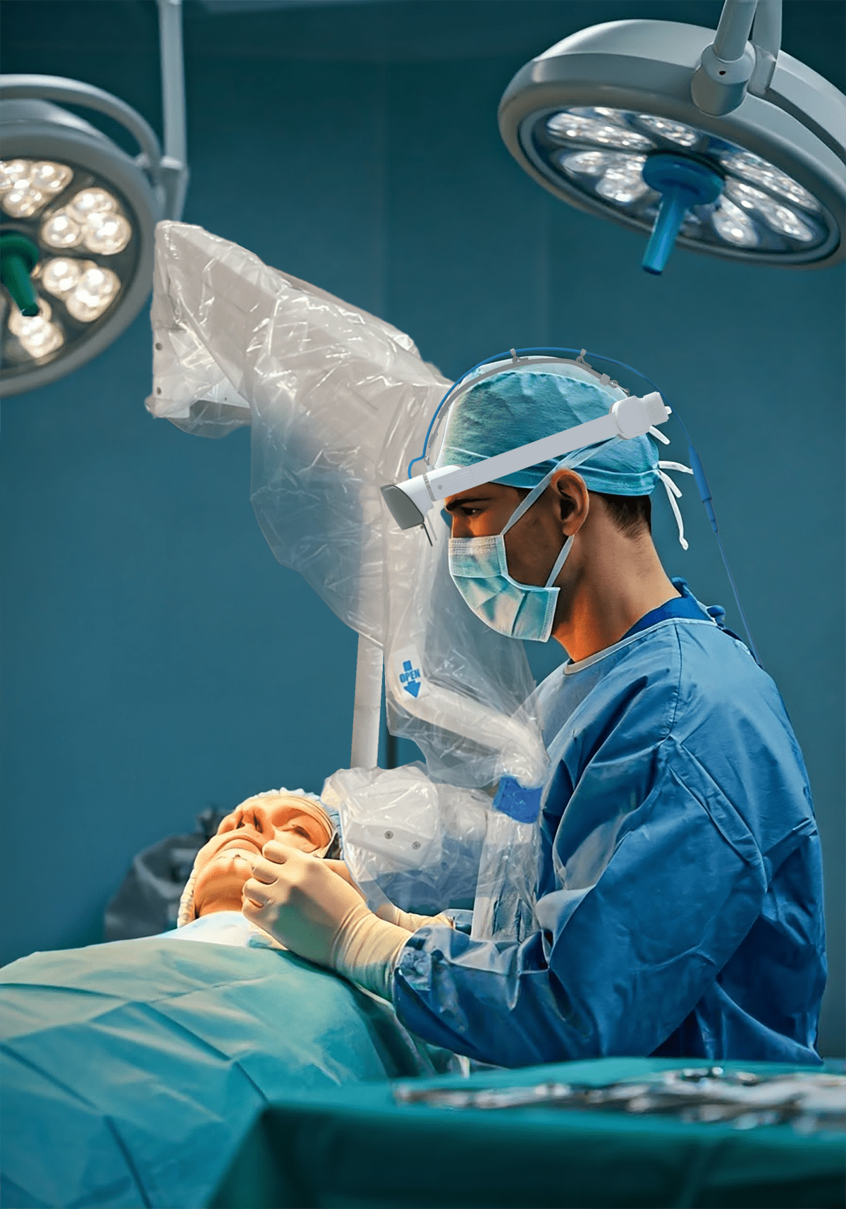
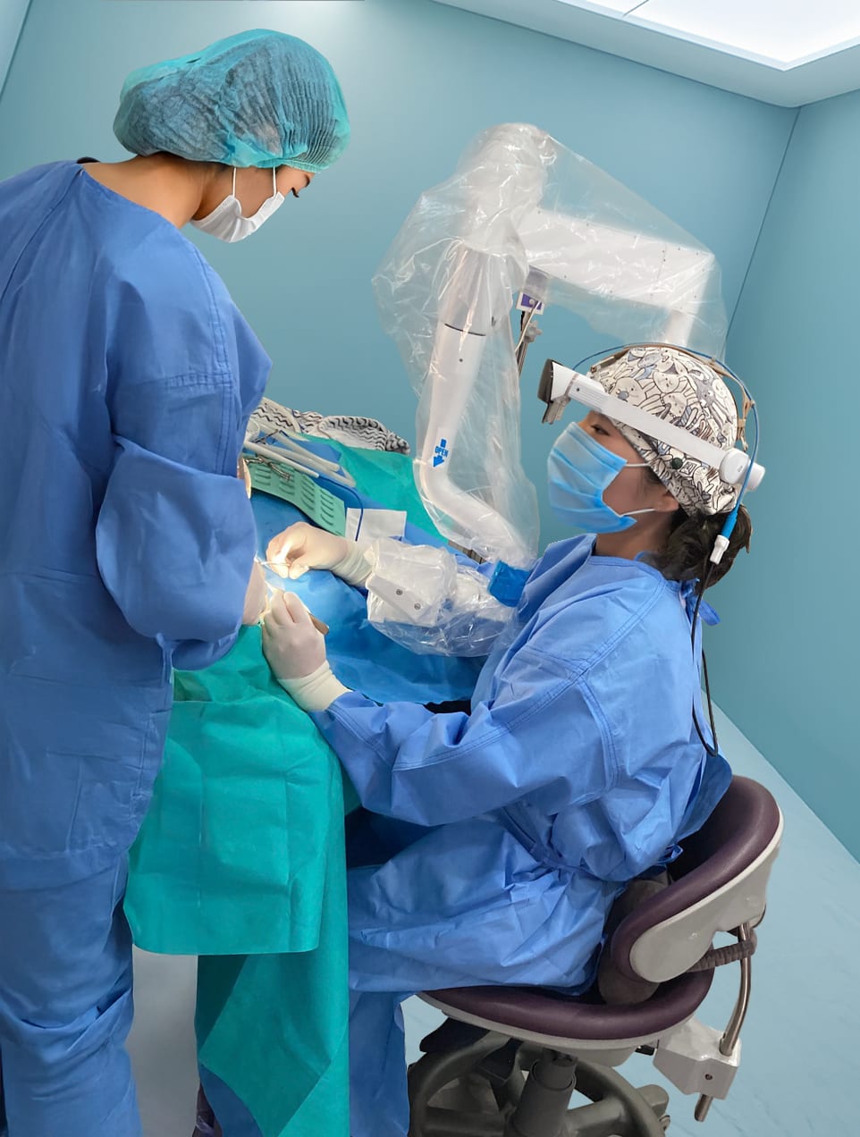
Traditionally, surgeons used loupes, looking down at the surgical site. With the 3D Microscope, there’s a shift in the approach. Surgeons can sit comfortably, viewing the surgery area three-dimensionally on a large monitor, akin to watching a 3D movie. This clear view and magnification allow for precise observation of tiny blood vessels and nerves, enhancing surgical focus and enabling more accurate and stable surgery.
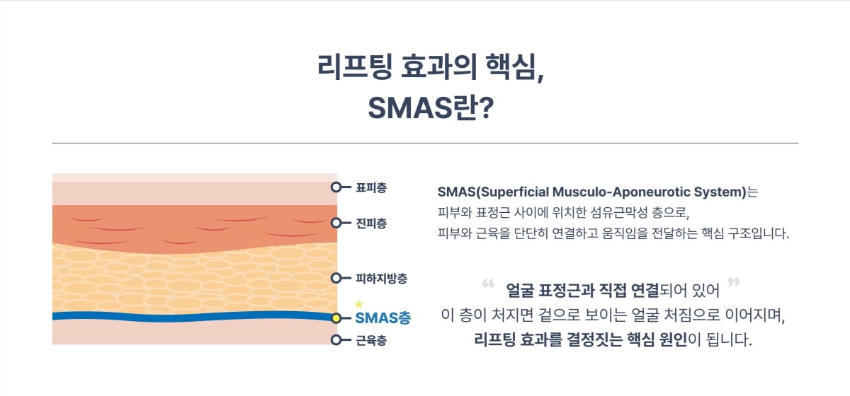
The Key to Natural Lifting: SMAS Layer Dissection
The focal point in facelift surgery is the SMAS, a fibromuscular layer just below the skin. With aging, this layer sags, causing wrinkles and looseness. Merely tightening the skin doesn’t offer a fundamental solution; accurately dissecting and securely repositioning the SMAS layer is crucial.
The challenge lies in the intricate network of facial expression-controlling nerves and blood vessels in this area. Nerve damage could result in unnatural expressions or sensory issues, thus requiring great caution. The 3D Microscope excels in this domain. By viewing in three dimensions, it allows for precise identification of nerves and blood vessels, minimizing tissue damage. It’s comparable to leisurely strolling a broad, well-lit path rather than navigating a narrow, dark road with a single flashlight.
The Real Difference Manifests Post-Surgery
“My friends say it doesn’t even look like I’ve had surgery, just that I’ve naturally gotten younger.”
Hearing such feedback from patients is incredibly rewarding. 3D Microscope Facelift transcends mere technological change, affecting both recovery and outcomes.
The precision in dissection decreases unnecessary tissue damage, thus reducing bruising and swelling. Quicker recovery facilitates a faster return to daily life. With maximum preservation of expression-controlling nerves, concerns about post-surgery awkward expressions are alleviated. Most importantly, the focus isn’t on forcibly tightening the skin but on naturally revitalizing facial contours by elevating the deeper layers properly. It isn’t about creating an artificial face but restoring one’s original appearance.

Choose Genuine Rejuvenation Wisely
While technology advances, its application remains in human hands. Entrusting your precious face to a surgery that reflects you in the mirror for years to come requires careful decision-making.
Real rejuvenation isn’t merely about erasing wrinkles but restoring a lively, healthy impression. What could natural youth, perfectly suited to your face, look like? Let’s consider it together.
Since the introduction of the 3D Microscope, facelift surgery precision and safety have been significantly enhanced. The broad, three-dimensional view allows for the identification of minute structures, ensuring positive changes even in the recovery process. For more professional details about the technology, contact us.
The subtle aspects unseen to the eye are promised to be handled excellently with the 3D Microscope for the best surgical outcomes.
Now, consider 3D SCOPEYE!
