
Is the 3D Microscope a Revolutionary Innovation in Medical Technology?
Stepping into the operating room equipped with a 3D microscope felt like entering a new world. The fresh visual experience this equipment offers to doctors is indeed revolutionary. Having worked nearly two decades as a plastic surgeon, addressing patients’ concerns, I’ve witnessed how changes in skin and facial features with aging, postpartum elasticity loss, and marks left after dieting become significant worries.
Many clients visiting the hospital with these concerns have particularly sensitive skin or complex vascular structures. “I break out no matter what I use…”, one client mentioned, which stayed with me. Such delicate needs make the limitations of conventional procedures more apparent. In the past, concerns about repeated treatments, short-lasting results, and side effects were always looming, and dissatisfaction with “awkwardness” or “noticeable changes” was a major worry for patients. However, recent medical technologies have evolved in a different direction. Instead of making significant changes, the focus is now on ‘precisely, bit by bit’ handling, which is gaining attention. The trend nowadays is adjusting specific areas subtly to achieve ‘naturalness without excess’. The 3D microscope is among the core technologies that make this precision possible.
What Makes the 3D Microscope Special?
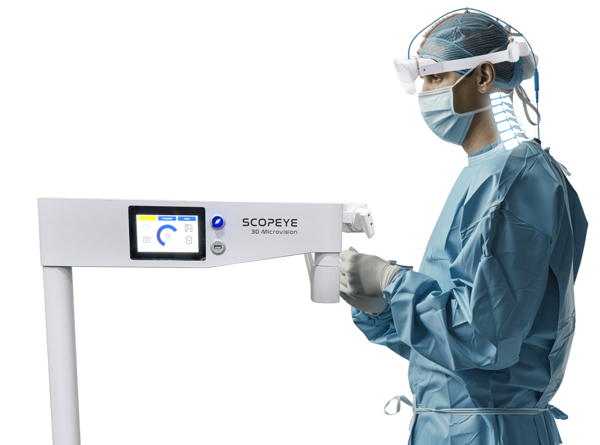
Visual clarity during surgery determines the success of the outcome, which is why the 3D microscope is garnering the attention of many doctors. This device provides a more precise view of the surgical area through depth-rich 3D images. This allows for easier differentiation of minute differences, enhancing surgical accuracy. By manifesting depth and dimensionality, which is hard to grasp with 2D flat images, it helps in better understanding the structure of the surgical area. For delicate surgeries, like those involving very thin layers of skin with minute blood vessels or nerve positions, it was difficult to distinguish precisely with the naked eye or traditional 2D microscopes. However, the 3D microscope displays these structures in a three-dimensional manner, allowing more realistic verification of the actual view.
“Even for the most delicate and sensitive area surgeries, this level of precise view is sufficient.”
This excellent visual information aids medical professionals in making more precise judgments and delicate maneuvers. Consequently, unnecessary tissue damage can be minimized, and the surrounding healthy tissue of the surgical area preserved as much as possible. This positively impacts the patient’s recovery speed and helps reduce the risk of potential post-surgery side effects. Beyond mere magnification, the 3D microscope contributes to enhancing the precision and safety of procedures by enabling a three-dimensional understanding and approach to the surgical space.
Why Do Experts Use the 3D Microscope?
One of the benefits of the 3D microscope is reduced fatigue and enhanced precision during surgeries. While traditional 2D images cause eye strain over prolonged use, the 3D microscope offers a more natural visual experience through its dimensional images, alleviating these issues. This allows for prolonged concentration during surgery. During long surgeries, there comes a point when concentration wanes. Bending over with loupes, dissecting, and repeating movements can stiffen the neck and ache the shoulders. I once performed surgeries that way, and I’d consistently massage my neck afterward. Although loupes were undoubtedly useful tools, the narrow field of view and limited depth perception made prolonged concentration challenging.
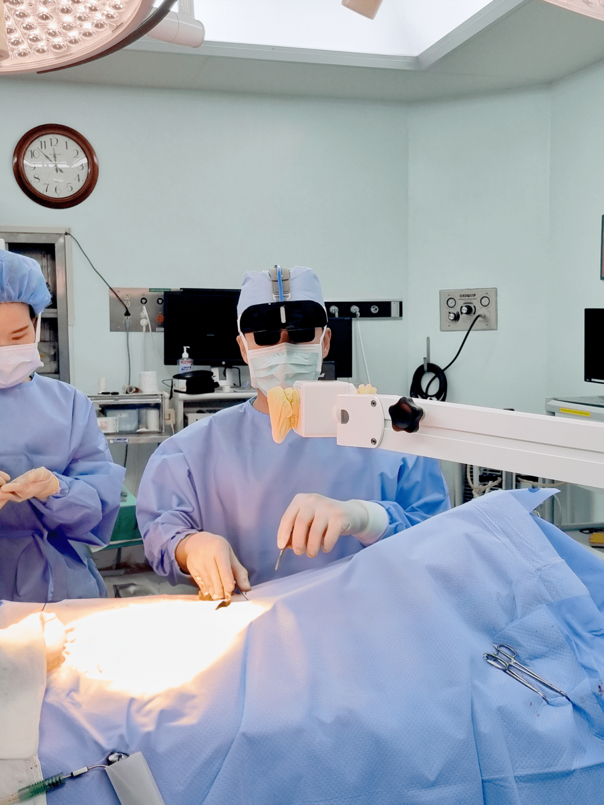
Yet, the 3D microscope allows medical professionals to observe the surgical area on a wide screen in a comfortable posture. Without the need to bend forward, one can watch straight ahead at a large monitor while doing the surgery, reducing physical strain. With no visual restrictions, it’s possible to view the entire surgical area at once and zoom in for detailed examination if necessary. This environment enhances the medical team’s focus, improving the quality of surgery. The better the surgeon’s condition, the more precise and stable the procedure becomes, which in turn provides safer and more predictable outcomes for patients. The observation power of the 3D scope, complementing loupes’ limitations, is evaluated as a tool that can elevate surgical precision.

How Is It Used in Plastic Surgery?
Recently, in some plastic surgeries, 3D microscopes are used to increase precision in facial skeletal or reconstructive procedures. Particularly in areas close to complex blood vessels or nerves, the role of this device can become crucial. This provides patients with safer and more meticulous surgical outcomes. In plastic surgery, the strength of the 3D microscope isn’t just about ‘seeing better’. For instance, during facial lifts or eye shape corrections, where fine nerves and blood vessels are densely packed, the 3D microscope can visualize structures not discernible to the naked eye more distinctly. This helps prevent unnecessary tissue damage and lower the risk of complications such as nerve damage or excessive bleeding.
This enhances the quality of plastic surgery and can positively influence the patient’s recovery process.
Which surgical environment would you prefer? Precise and stress-free recovery, which would be your more trustworthy standard?

Recently in cosmetic procedures, the focus is on how naturally changes occur without being excessive. In this trend, 3D microscope technology is becoming a meaningful turning point in plastic surgery by pushing past visual limitations to broaden the possibilities for precise procedures. If you have further inquiries, please contact us.
3D microscopes guarantee the best surgical results with precision that is invisible to the naked eye.
Now, try 3D SCOPEYE!
